What if a birthmark wasn't just a small, innocuous spot, but a vast, disfiguring lesion casting a long shadow on a child's life? The reality of giant congenital hairy nevus is a stark reminder that some are born carrying burdens far heavier than others, demanding innovative medical interventions and unwavering emotional support.
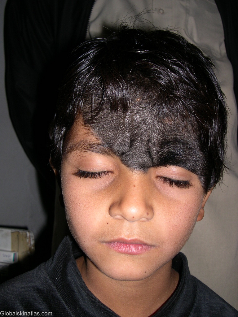
Consider the case of a 9-year-old boy from Nepal who recently presented at the outpatient department of a reconstructive surgery center. His condition: a giant pigmented hairy nevus of the face. The lesion was not confined to a small area; it aggressively occupied the right zygomatic region, the right cheek, the right upper and lower eyelids, the right eyebrow, and the entirety of his nose, even extending onto the left infraorbital region. Its dimensions were striking, measuring a full 12 centimeters. This wasn't merely a cosmetic issue; it was a significant medical challenge with potential long-term implications.
| Category | Details |
|---|---|
| Patient Information | |
| Age | 9 years old |
| Origin | Nepal |
| Medical Condition | |
| Diagnosis | Giant Pigmented Hairy Nevus of the Face |
| Location of Lesion | Right zygomatic region, right cheek, right upper and lower eyelids, right eyebrow, nose (extending to left infraorbital region) |
| Size of Lesion | 12 cm |
| Nevus Information | |
| Alternative Names | Giant Congenital Melanocytic Nevus, Bathing Trunk Nevus, Garment Nevus, Nevus Pigmentosus et Pilosus |
| Characteristics | Large, darkly pigmented, often hairy patches present at birth |
| Potential Risks | Increased risk of melanoma (skin cancer) |
| Treatment Options | Surgical removal, curettage, dermabrasion |
| Reference Website | National Institutes of Health (NIH) - Congenital Melanocytic Nevus |
A hairy nevus that attains or surpasses 8 inches in diameter by adulthood is classified as a giant congenital hairy nevus. It is imperative to understand that while larger hairy nevi carry a heightened risk of skin cancer, hasty conclusions should be avoided until a qualified medical professional has thoroughly examined and diagnosed the nevi. Premature assumptions can lead to unnecessary anxiety and potentially misdirected treatment strategies.
- Kaitlyn Frohnapfel Drew Mcintyres Wife Her Life Career
- John Mallory Asher Weird Science Star Director More
These lesions typically manifest as a singular entity, often situated on the scalp or the face, and are present from birth. This distinguishes them from other types of nevi, such as nevus sebaceous, which presents as a circumscribed, slightly elevated, hairless plaque, usually lacking the pigmentation characteristic of a congenital melanocytic nevus (CMN). During puberty, a nevus sebaceous undergoes transformation, becoming verrucous and nodular, sometimes exhibiting areas of linear distribution. This difference in presentation is crucial for accurate diagnosis and management.
The prophylactic surgical removal of a naevus is a consideration that requires careful deliberation. Several factors must be meticulously evaluated before embarking on such a course of action. The size and location of the nevus, the age and overall health of the patient, and the potential for scarring and other complications all play a role in the decision-making process. A comprehensive assessment ensures that the benefits of removal outweigh the risks.
A congenital melanocytic nevus (CMN) is fundamentally a benign skin lesion stemming from the proliferation of nevomelanocytes. It is evident either at birth or within the initial weeks of life. CMNs are typically categorized based on size: small, medium, large, or giant. This classification is not arbitrary; it directly correlates with the potential for malignant transformation. Larger CMNs inherently pose a greater risk of developing into melanoma later in life.
- Belle Gibsons Son Truth About Her Lies Motherhood Exposed
- Hilary Duffs Bold Nude Photoshoot Body Positivity More
While these moles are commonly observed on the trunk or limbs, they possess the capacity to appear anywhere on the body's surface. The presence of excess hair growth is a frequent characteristic, leading to these lesions sometimes being referred to as giant hairy nevi. This terminology is descriptive but should not overshadow the underlying medical significance of the condition.
Although giant congenital nevi can arise at any anatomical site, a predilection exists for their occurrence on the trunk, encompassing the back, abdomen, hips, and buttocks. The scalp and face are also frequently involved, as illustrated by the case of the young boy from Nepal. The location of the nevus significantly influences treatment strategies, as some areas are more amenable to surgical intervention than others.
In essence, a congenital hairy nevus, or congenital nevus, is a large mole present at birth. The underlying cause lies in the hyperactivity of pigment-producing cells, which impart color to the skin. This hyperactivity results in an overabundance of melanin, leading to the formation of the nevus. Congenital nevi exhibit a wide spectrum of sizes and shapes, ranging from small, barely noticeable marks to expansive lesions covering significant portions of the body.
One approach to managing extensive hairy nevi involves curettage or dermabrasion during the initial weeks of life. This technique aims to remove the superficial layers of the nevus, reducing pigmentation and hair growth. However, it is crucial to recognize that recurrence of pigmentation and hair is possible, necessitating long-term follow-up. The success of curettage is contingent on factors such as the depth of the nevus and the individual's healing response.
Most moles are benign, posing no threat to health. However, the presence of hair within a mole can be a source of concern for some individuals. Despite the popular myth linking hairy moles to cancer, this association is largely unfounded. While any mole exhibiting changes in size, shape, or color should be evaluated by a dermatologist, the presence of hair alone is not indicative of malignancy.
While most congenital nevi do not precipitate health problems, a small percentage harbors the potential to develop into skin cancer, specifically melanoma, later in life. The likelihood of melanoma increases in direct proportion to the size of the nevus. Regular monitoring and sun protection are essential for individuals with congenital nevi, particularly those classified as large or giant.
A large congenital melanocytic nevus (LCMN) represents a rare form of congenital mole, occurring in approximately 1 in 20,000 newborns globally. This rarity underscores the need for specialized medical expertise in managing these cases. LCMNs often present unique challenges due to their size, location, and potential for complications.
The scalp's skin, being relatively tight, often poses challenges in the complete surgical removal of a nevus in a single procedure. Insufficient skin elasticity may necessitate staged excisions or other reconstructive techniques. A carefully planned surgical approach is crucial to achieving optimal cosmetic and functional outcomes.
In one instance, a young patient underwent her initial surgery with the Little Baby Face Foundation in November 2022. The primary goal of this procedure was to remove the majority of the hairy nevus, demonstrating the commitment of specialized organizations to addressing these complex cases. Such interventions can significantly improve a child's quality of life, both physically and emotionally.
Following curettage of hairy nevi in the immediate aftermath of birth, the recurrence of pigmentation and hair growth remains a possibility on long-term follow-up. This highlights the importance of continuous monitoring and potential need for additional interventions. Parents and caregivers should be educated about the signs of recurrence and the importance of adherence to follow-up appointments.
The management of giant hairy nevi is a multifaceted endeavor, contingent on various factors, including the size and anatomical location of the lesion. A comprehensive approach is essential, encompassing surgical expertise, reconstructive techniques, and psychological support. Each patient presents a unique set of challenges, demanding individualized treatment plans.
A giant congenital melanocytic nevus, also known as bathing trunk nevus, garment nevus, giant hairy nevus, or nevus pigmentosus et pilosus, is characterized by one or more large, darkly pigmented, and sometimes hairy patches. The terms "bathing trunk nevus" and "garment nevus" aptly describe the lesion's extensive coverage, often resembling the area covered by these articles of clothing.
These lesions carry a relatively elevated risk of malignant transformation, necessitating vigilant monitoring and proactive management. The potential for melanoma development underscores the importance of early detection and intervention. Regular self-examinations and professional skin checks are crucial for individuals with giant congenital nevi.
Giant congenital nevi frequently manifest as dark brown to black patches of skin, covering areas such as the trunk, neck, back, limbs, or face. These patches typically exhibit a defined, singular border and a surface area characterized by hair and a markedly darker pigmentation compared to the surrounding skin. This pronounced contrast makes the lesions easily distinguishable.
Nevi, the medical term for moles, are common skin lesions. Most individuals have between 10 and 40 moles. These common nevi are typically harmless collections of colored cells, appearing as small, well-defined spots on the skin. However, congenital nevi, particularly those classified as large or giant, require specialized attention due to their potential for complications.
Pigmented hairy epidermal nevus of the face is a specific presentation of this condition, emphasizing the combined characteristics of pigmentation, hair growth, and epidermal involvement. This type of nevus can pose significant cosmetic and functional challenges, particularly when located on the face.
The presence of hair in a CMN can be evident at birth or develop over time. The growth of hair within the nevus is attributed to the proliferation of hair follicles within the lesion. This characteristic, while not inherently dangerous, can contribute to the overall cosmetic impact of the nevus.
Patients can continue to develop new, smaller CMNs over time, with significant variability between individuals. This phenomenon underscores the importance of ongoing monitoring, even after initial treatment. The appearance of new nevi should be promptly evaluated by a dermatologist.
The older term "neurocutaneous melanosis" refers to the association of CMN on the skin with a subset of neurological abnormalities. This association highlights the potential for systemic involvement in some cases of CMN, necessitating a multidisciplinary approach to management.
The congenital hairy nevus fundamentally represents a pigmented surface lesion present at birth. Its defining characteristics are its presence from birth, its pigmentation, and the presence of hair. These features distinguish it from other types of skin lesions that may develop later in life.
Giant hairy nevi assume particular significance due to their predisposition to malignant transformation. This potential for melanoma development necessitates vigilant monitoring and proactive management strategies. Early detection and intervention are crucial for improving outcomes.
Surgical removal represents the most common treatment modality for hairy nevi. The goal of surgical removal is to excise the nevus completely, minimizing the risk of recurrence and malignant transformation. The specific surgical technique employed depends on the size, location, and depth of the nevus.
Removal of smaller nevi can typically be accomplished in a single procedure. However, larger nevi may necessitate staged excisions or reconstructive techniques to achieve optimal cosmetic and functional outcomes. The surgical approach should be carefully tailored to the individual patient's needs.
Congenital melanocytic nevus of the skin of the lower eyelid, epidermal nevus of the left lower eyelid, hyperpigmentation of the left eyelid and periocular area, melanocytic nevus of the left lower eyelid these are all specific examples of how CMNs can manifest in the delicate area around the eyes. Such lesions can pose significant cosmetic and functional challenges, potentially affecting vision and eyelid function.
Excessive hair growth, or hypertrichosis, can occur within the nevus. This is attributed to the proliferation of hair follicles within the lesion. While not inherently dangerous, hypertrichosis can contribute to the overall cosmetic impact of the nevus.
There is often less fat tissue beneath the skin of the nevus. This can result in a thinner appearance of the skin in the affected area compared to other parts of the body. The lack of subcutaneous fat can also make the nevus more susceptible to injury and irritation.
Individuals with giant congenital melanocytic nevus may have multiple nevi, with the other nevi often being smaller than the giant nevus. This phenomenon underscores the importance of a comprehensive skin examination to identify and monitor all nevi present. Each nevus should be evaluated for its potential for malignant transformation.
- Madalina Cojocari What Happened Latest Updates Timeline
- Pearl Carter From Youngest Pilot To Backstreet Baby Names